BAP31 Recombinant Rabbit Monoclonal Antibody [JG37-81]

Catalog# ET7108-54
BAP31 Recombinant Rabbit Monoclonal Antibody [JG37-81]
-
WB
-
IF-Cell
-
IF-Tissue
-
IHC-P
-
FC
-
Human
-
Mouse
概述
产品名称
BAP31 Recombinant Rabbit Monoclonal Antibody [JG37-81]
抗体类型
Recombinant Rabbit monoclonal Antibody
免疫原
Recombinant protein within Human BAP31 aa 81-220 / 246.
种属反应性
Human, Mouse
验证应用
WB, IF-Cell, IF-Tissue, IHC-P, FC
分子量
28 kDa
阳性对照
A431 cell lysate, SK-Br-3 cell lysate, A431, A549, PC-3M, human liver tissue, human colon carcinoma tissue, human kidney tissue, mouse testis tissue, Hela.
偶联
unconjugated
克隆号
JG37-81
RRID
产品特性
形态
Liquid
浓度
1ug/ul
存放说明
Store at +4℃ after thawing. Aliquot store at -20℃. Avoid repeated freeze / thaw cycles.
存储缓冲液
1*TBS (pH7.4), 0.05% BSA, 40% Glycerol. Preservative: 0.05% Sodium Azide.
亚型
IgG
纯化方式
Protein A affinity purified.
应用稀释度
-
WB
-
1:500-1:2,000
-
IF-Cell
-
1:50-1:200
-
IHC-P
-
1:50-1:200
-
FC
-
1:50-1:100
靶点
功能
BAP31, a human Bcl-2-interacting protein, is an integral membrane protein that is a component of a protein complex in the endoplasmic reticulum. This protein complex mechanically bridges an apoptosis-initiating caspase, like procaspase-8, with the anti-apoptotic regulator Bcl-2 or Bcl-XL. The cytosolic domain of BAP31 contains two identical caspase recognition sites, which are preferentially cleaved by initiator caspases, including caspase 8. Cleavage of BAP31 during apoptosis generates a p20 fragment, which remains integrated in the membrane and, when expressed ectopically, is a potent inducer of cell death. BAP31 cleavage is important for manifesting cytoplasmic apoptotic events associated with membrane fragmentation and in the cross talk between mitochondria and the endoplasmic reticulum during Fas- mediated apoptosis. The BAP31 gene is ubiquitously expressed in murine tissues and is located on the X chromosome in both mouse and human.
背景文献
1. Annaert W G et al. Export of cellubrevin from the endoplasmic reticulum is controlled by BAP31. J Cell Biol 139:1397-1410 (1997).
2. Paquet M E et al. Bap29/31 influences the intracellular traffic of MHC class I molecules. J Immunol 172:7548-7555 (2004).
序列相似性
Belongs to the BCAP29/BCAP31 family.
组织特异性
Ubiquitous. Highly expressed in neurons and discrete endocrine cells.
翻译后修饰
Cleaved by CASP8 and other caspases.
亚细胞定位
Endoplasmic reticulum membrane.
UNIPROT #
别名
6C6 AG antibody
6C6 AG tumor associated antigen antibody
6C6-AG tumor-associated antigen antibody
6C6AG antibody
6C6AG tumor associated antigen antibody
Accessory protein BAP 31 antibody
Accessory protein BAP31 antibody
B cell receptor associated protein 31 antibody
B-cell receptor-associated protein 31 antibody
BA31 antibody
展开图片
-
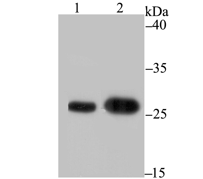
Western blot analysis of BAP31 on different lysates. Proteins were transferred to a PVDF membrane and blocked with 5% BSA in PBS for 1 hour at room temperature. The primary antibody (ET7108-54, 1/500) was used in 5% BSA at room temperature for 2 hours. Goat Anti-Rabbit IgG - HRP Secondary Antibody (HA1001) at 1:200,000 dilution was used for 1 hour at room temperature.
Positive control:
Lane 1: A431 cell lysate
Lane 2: SK-Br-3 cell lysate -
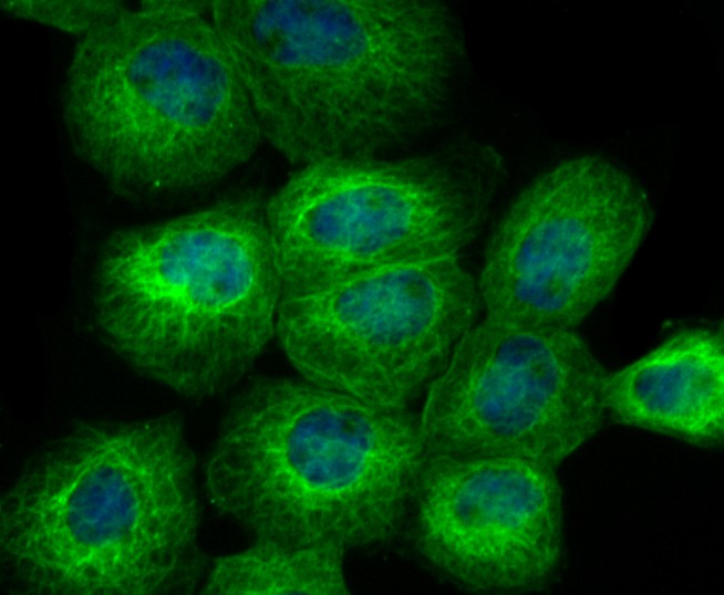
ICC staining of BAP31 in A431 cells (green). Formalin fixed cells were permeabilized with 0.1% Triton X-100 in TBS for 10 minutes at room temperature and blocked with 1% Blocker BSA for 15 minutes at room temperature. Cells were probed with the primary antibody (ET7108-54, 1/50) for 1 hour at room temperature, washed with PBS. Alexa Fluor®488 Goat anti-Rabbit IgG was used as the secondary antibody at 1/1,000 dilution. The nuclear counter stain is DAPI (blue).
-
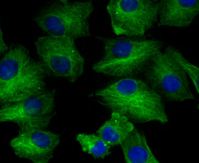
ICC staining of BAP31 in A549 cells (green). Formalin fixed cells were permeabilized with 0.1% Triton X-100 in TBS for 10 minutes at room temperature and blocked with 1% Blocker BSA for 15 minutes at room temperature. Cells were probed with the primary antibody (ET7108-54, 1/50) for 1 hour at room temperature, washed with PBS. Alexa Fluor®488 Goat anti-Rabbit IgG was used as the secondary antibody at 1/1,000 dilution. The nuclear counter stain is DAPI (blue).
-
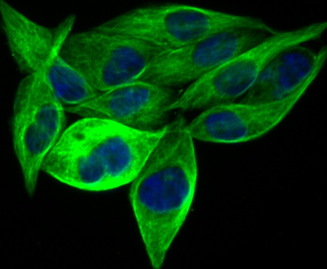
ICC staining of BAP31 in PC-3M cells (green). Formalin fixed cells were permeabilized with 0.1% Triton X-100 in TBS for 10 minutes at room temperature and blocked with 1% Blocker BSA for 15 minutes at room temperature. Cells were probed with the primary antibody (ET7108-54, 1/50) for 1 hour at room temperature, washed with PBS. Alexa Fluor®488 Goat anti-Rabbit IgG was used as the secondary antibody at 1/1,000 dilution. The nuclear counter stain is DAPI (blue).
-
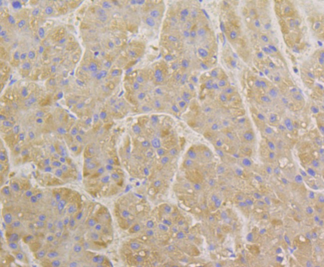
Immunohistochemical analysis of paraffin-embedded human liver tissue using anti-BAP31 antibody. The section was pre-treated using heat mediated antigen retrieval with Tris-EDTA buffer (pH 8.0-8.4) for 20 minutes.The tissues were blocked in 5% BSA for 30 minutes at room temperature, washed with ddH2O and PBS, and then probed with the primary antibody (ET7108-54, 1/50) for 30 minutes at room temperature. The detection was performed using an HRP conjugated compact polymer system. DAB was used as the chromogen. Tissues were counterstained with hematoxylin and mounted with DPX.
-
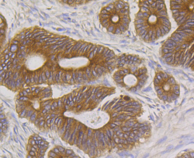
Immunohistochemical analysis of paraffin-embedded human colon carcinoma tissue using anti-BAP31 antibody. The section was pre-treated using heat mediated antigen retrieval with Tris-EDTA buffer (pH 8.0-8.4) for 20 minutes.The tissues were blocked in 5% BSA for 30 minutes at room temperature, washed with ddH2O and PBS, and then probed with the primary antibody (ET7108-54, 1/50) for 30 minutes at room temperature. The detection was performed using an HRP conjugated compact polymer system. DAB was used as the chromogen. Tissues were counterstained with hematoxylin and mounted with DPX.
-
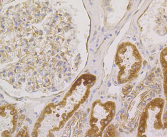
Immunohistochemical analysis of paraffin-embedded human kidney tissue using anti-BAP31 antibody. The section was pre-treated using heat mediated antigen retrieval with Tris-EDTA buffer (pH 8.0-8.4) for 20 minutes.The tissues were blocked in 5% BSA for 30 minutes at room temperature, washed with ddH2O and PBS, and then probed with the primary antibody (ET7108-54, 1/50) for 30 minutes at room temperature. The detection was performed using an HRP conjugated compact polymer system. DAB was used as the chromogen. Tissues were counterstained with hematoxylin and mounted with DPX.
-
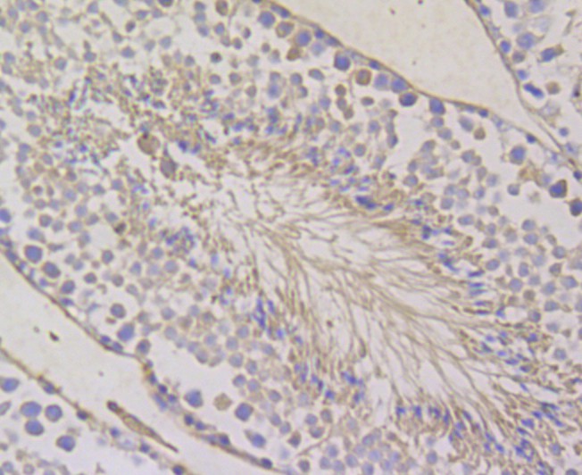
Immunohistochemical analysis of paraffin-embedded mouse testis tissue using anti-BAP31 antibody. The section was pre-treated using heat mediated antigen retrieval with Tris-EDTA buffer (pH 8.0-8.4) for 20 minutes.The tissues were blocked in 5% BSA for 30 minutes at room temperature, washed with ddH2O and PBS, and then probed with the primary antibody (ET7108-54, 1/50) for 30 minutes at room temperature. The detection was performed using an HRP conjugated compact polymer system. DAB was used as the chromogen. Tissues were counterstained with hematoxylin and mounted with DPX.
-
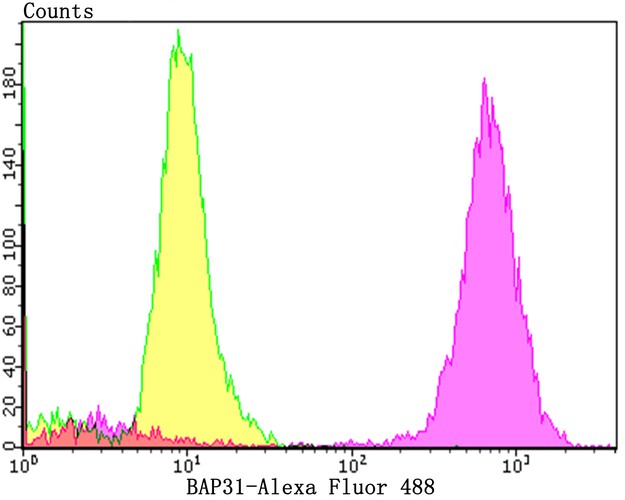
Flow cytometric analysis of BAP31 was done on Hela cells. The cells were fixed, permeabilized and stained with the primary antibody (ET7108-54, 1/50) (purple). After incubation of the primary antibody at room temperature for an hour, the cells were stained with a Alexa Fluor 488-conjugated Goat anti-Rabbit IgG Secondary antibody at 1/1000 dilution for 30 minutes.Unlabelled sample was used as a control (cells without incubation with primary antibody; yellow).
Please note: All products are "FOR RESEARCH USE ONLY AND ARE NOT INTENDED FOR DIAGNOSTIC OR THERAPEUTIC USE"