Transferrin Receptor 2 Recombinant Rabbit Monoclonal Antibody [JG58-34]


Catalog# ET7108-21
Transferrin Receptor 2 Recombinant Rabbit Monoclonal Antibody [JG58-34]
-
WB
-
IHC-P
-
Human
概述
产品名称
Transferrin Receptor 2 Recombinant Rabbit Monoclonal Antibody [JG58-34]
抗体类型
Recombinant Rabbit monoclonal Antibody
免疫原
Recombinant protein within Human Transferrin Receptor 2 aa 130-320 / 801.
种属反应性
Human
验证应用
WB, IHC-P
分子量
Predicted band size: 89 kDa
阳性对照
K562 cell lysates, human liver tissue, human liver carcinoma tissue.
偶联
unconjugated
克隆号
JG58-34
RRID
产品特性
形态
Liquid
浓度
1ug/ul
存放说明
Store at +4℃ after thawing. Aliquot store at -20℃. Avoid repeated freeze / thaw cycles.
存储缓冲液
1*TBS (pH7.4), 0.05% BSA, 40% Glycerol. Preservative: 0.05% Sodium Azide.
亚型
IgG
纯化方式
Protein A affinity purified.
应用稀释度
-
WB
-
1:500-1:2,000
-
IHC-P
-
1:50-1:1,000
靶点
功能
Iron is a vital molecule for living organisms because it is involved in a wide variety of metabolic processes, such as oxygen transport, DNA synthesis and electron transport. Excessive iron uptake leads to tissue damage as a result of formation of free radicals. Iron uptake and storage is tightly regulated by the feedback system of iron responsive element-containing gene products and iron regulatory proteins that modulate the expression levels of the genes involved in iron metabolism. The transferrin receptor 2 (TFR2) mediates the uptake of transferrin-bound iron. It is involved in iron metabolism, hepatocyte function and erythrocyte differentiation, and is highly expressed as a protein in liver as well as in hepatocytes and erythroid precursors. The gene encoding human TRF2 maps to chromosome 7q22 and is expressed as an a isoform, which encodes a transmembrane protein, and a b isoform, which encodes a shorter, intracellular protein. Mutations in the TFR2 gene result in hereditary hemochromatosis type III (HFE3), an iron overloading disorder that results in clinical complications, including cirrhosis, cardiopathy, diabetes, endocrine dysfunctions, arthropathy and susceptibility to liver cancer.
背景文献
1. Roetto A et al. New mutations inactivating transferrin 2 in hemochromatosis type 3. Blood 97:2555-2560 (2001).
2. Mattman A et al. Transferrin receptor 2 (TfR2) and HFE mutational analysis in non-C282Y iron overload: identification of a novel TfR2 mutation. Blood 100:1075-1077 (2002).
序列相似性
Belongs to the peptidase M28 family. M28B subfamily.
组织特异性
Predominantly expressed in liver. While the alpha form is also expressed in spleen, lung, muscle, prostate and peripheral blood mononuclear cells, the beta form is expressed in all tissues tested, albeit weakly.
亚细胞定位
Cell membrane. Cytoplasm.
UNIPROT #
别名
HFE 3 antibody
HFE3 antibody
MGC126368 antibody
TFR 2 antibody
TfR2 antibody
TFR2_HUMAN antibody
TFRC 2 antibody
TFRC2 antibody
Transferrin receptor 2 antibody
Transferrin receptor protein 2 antibody
图片
-
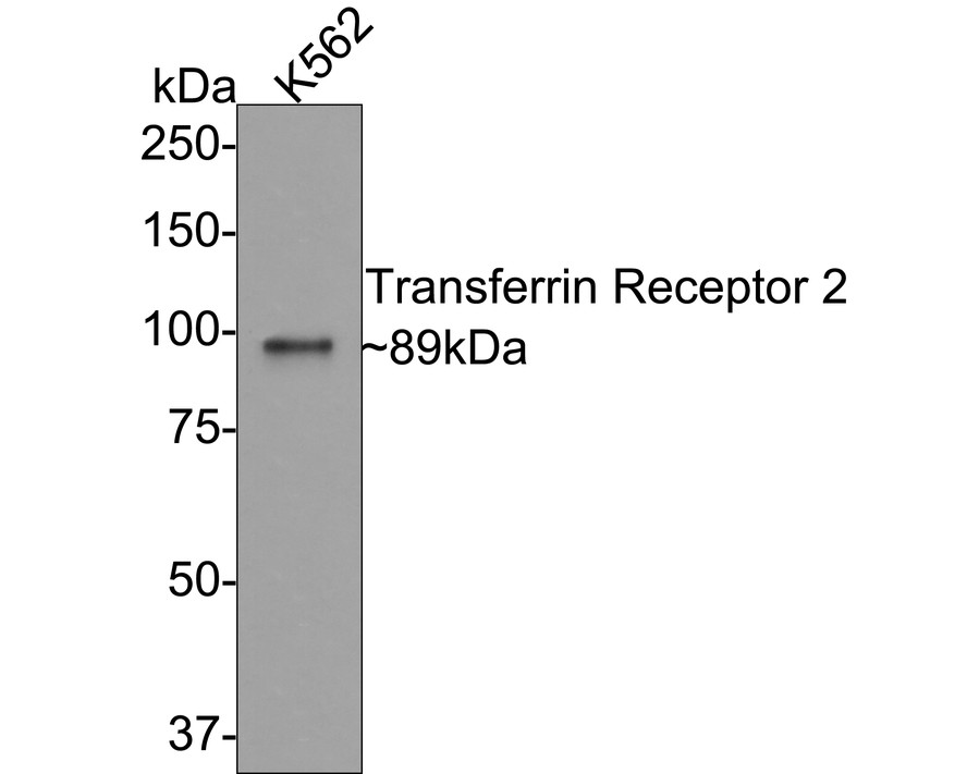
Western blot analysis of Transferrin Receptor 2 on K562 cell lysates with Rabbit anti-Transferrin Receptor 2 antibody (ET7108-21) at 1/500 dilution.
Lysates/proteins at 10 µg/Lane.
Predicted band size: 89 kDa
Observed band size: 89 kDa
Exposure time: 2 minutes;
8% SDS-PAGE gel.
Proteins were transferred to a PVDF membrane and blocked with 5% NFDM/TBST for 1 hour at room temperature. The primary antibody (ET7108-21) at 1/500 dilution was used in 5% NFDM/TBST at room temperature for 2 hours. Goat Anti-Rabbit IgG - HRP Secondary Antibody (HA1001) at 1:300,000 dilution was used for 1 hour at room temperature. -
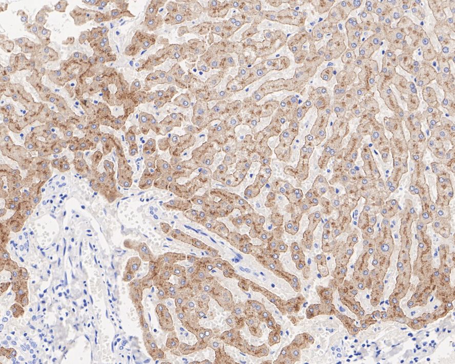
Immunohistochemical analysis of paraffin-embedded human liver tissue with Rabbit anti-Transferrin Receptor 2 antibody (ET7108-21) at 1/1,000 dilution.
The section was pre-treated using heat mediated antigen retrieval with Tris-EDTA buffer (pH 9.0) for 20 minutes. The tissues were blocked in 1% BSA for 20 minutes at room temperature, washed with ddH2O and PBS, and then probed with the primary antibody (ET7108-21) at 1/1,000 dilution for 1 hour at room temperature. The detection was performed using an HRP conjugated compact polymer system. DAB was used as the chromogen. Tissues were counterstained with hematoxylin and mounted with DPX. -
Immunohistochemical analysis of paraffin-embedded human liver carcinoma tissue with Rabbit anti-Transferrin Receptor 2 antibody (ET7108-21) at 1/1,000 dilution.
The section was pre-treated using heat mediated antigen retrieval with Tris-EDTA buffer (pH 9.0) for 20 minutes. The tissues were blocked in 1% BSA for 20 minutes at room temperature, washed with ddH2O and PBS, and then probed with the primary antibody (ET7108-21) at 1/1,000 dilution for 1 hour at room temperature. The detection was performed using an HRP conjugated compact polymer system. DAB was used as the chromogen. Tissues were counterstained with hematoxylin and mounted with DPX.
Please note: All products are "FOR RESEARCH USE ONLY AND ARE NOT INTENDED FOR DIAGNOSTIC OR THERAPEUTIC USE"