CD73 Mouse Monoclonal Antibody [A6F9]


Catalog# HA601011
CD73 Mouse Monoclonal Antibody [A6F9]
-
WB
-
IHC-P
-
IF-Tissue
-
FC
-
Human
概述
产品名称
CD73 Mouse Monoclonal Antibody [A6F9]
抗体类型
Mouse Monoclonal Antibody
免疫原
Recombinant protein within Human CD73 27-549.
种属反应性
Human
验证应用
WB, IHC-P, IF-Tissue, FC
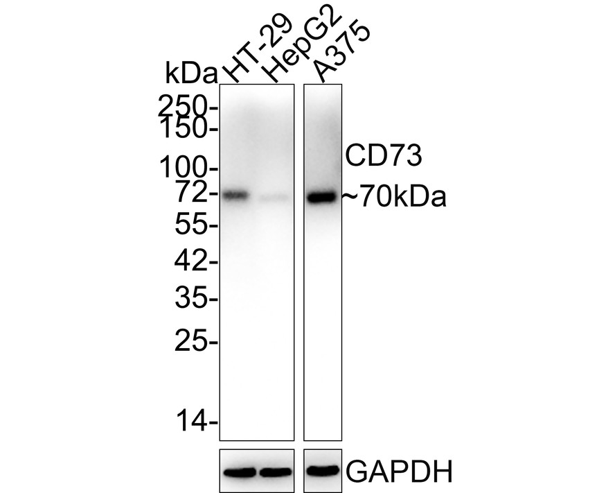
分子量
Predicted band size: 63 kDa
阳性对照
U87MG cell lysates, NCI-H441 xenograft tissue, U87MG xenograft tissue, U87-MG.
偶联
unconjugated
克隆号
A6F9
RRID
产品特性
形态
Liquid
浓度
1ug/ul
存放说明
Store at +4℃ after thawing. Aliquot store at -20℃. Avoid repeated freeze / thaw cycles.
存储缓冲液
PBS (pH7.4), 0.1% BSA, 40% Glycerol. Preservative: 0.05% Sodium Azide.
亚型
IgG1
纯化方式
Protein G affinity purified.
应用稀释度
-
WB
-
1:500
-
IHC-P
-
1:200-1:600
-
IF-Tissue
-
1:100
-
FC
-
1:500-1:1,000
靶点
功能
5′-nucleotidase (5′-NT), also known as ecto-5′-nucleotidase or CD73 (cluster of differentiation 73), is an enzyme that in humans is encoded by the NT5E gene. CD73 commonly serves to convert AMP to adenosine. Ecto-5-prime-nucleotidase catalyzes the conversion at neutral pH of purine 5-prime mononucleotides to nucleosides, the preferred substrate being AMP. The enzyme consists of a dimer of 2 identical 70-kD subunits bound by a glycosyl phosphatidyl inositol linkage to the external face of the plasma membrane. The enzyme is used as a marker of lymphocyte differentiation. Consequently, a deficiency of NT5 occurs in a variety of immunodeficiency diseases. Other forms of 5-prime nucleotidase exist in the cytoplasm and lysosomes and can be distinguished from ecto-NT5 by their substrate affinities, requirement for divalent magnesium ion, activation by ATP, and inhibition by inorganic phosphate. Rare allelic variants are associated with a syndrome of adult-onset calcification of joints and arteries (CALJA) affecting the iliac, femoral, and tibial arteries reducing circulation in the legs and the joints of the hands and feet causing pain. NT5E can act as an immune inhibitory control molecule. Free adenosine generated by NT5E inhibits cellular immune responses and thereby promotes immune escape of tumor cells.
背景文献
1. Harvey JB. et. al. CD73\'s Potential as an Immunotherapy Target in Gastrointestinal Cancers. Front Immunol. 2020 Apr
2. Neo SY. et. al. CD73 immune checkpoint defines regulatory NK cells within the tumor microenvironment. J Clin Invest. 2020 Mar
亚细胞定位
Cell membrane.
UNIPROT #
别名
5' NT antibody
5' nucleotidase (CD73) antibody
5' nucleotidase precursor antibody
5' nucleotidase, ecto antibody
5' nucleotidase, ecto (CD73) antibody
5'-NT antibody
5'-nucleotidase antibody
5NTD_HUMAN antibody
CD73 antibody
CD73 antigen antibody
展开图片
-
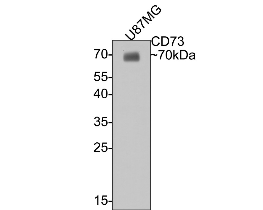
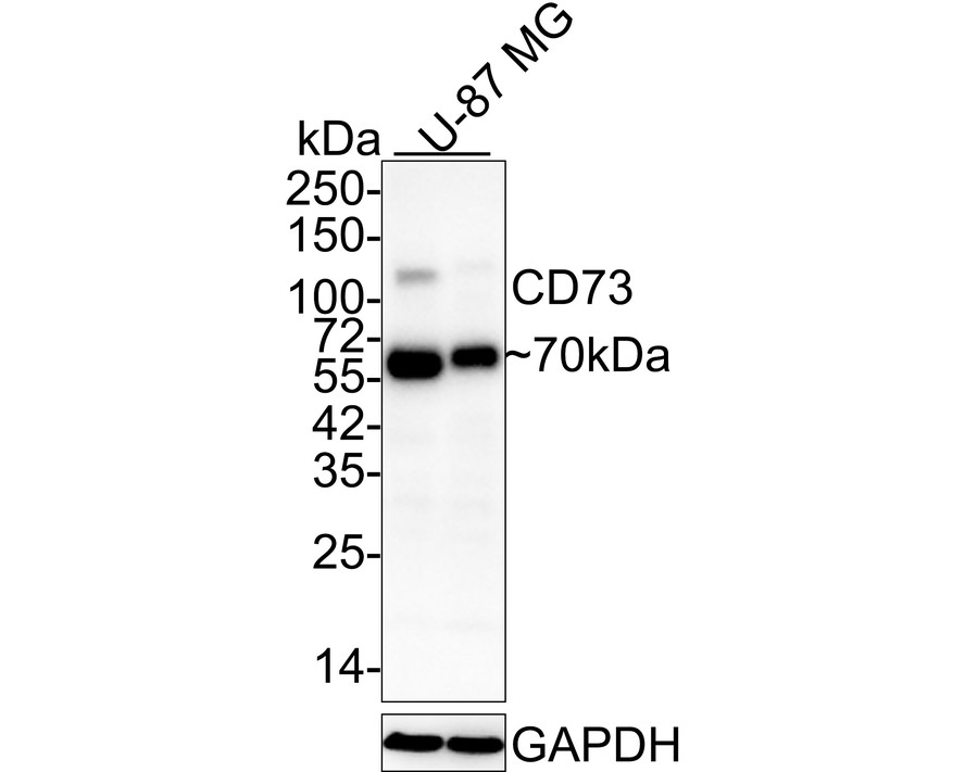
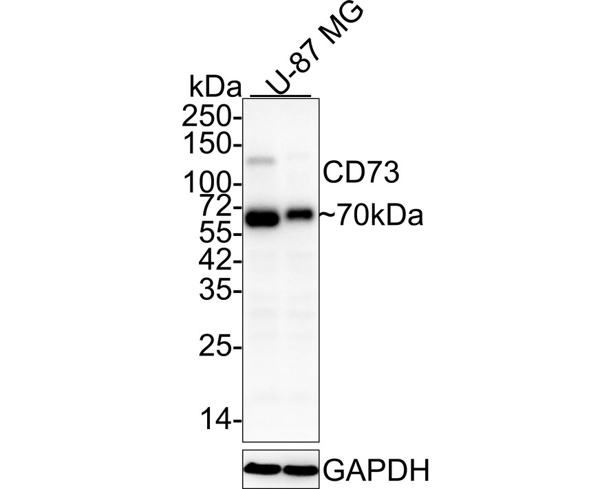
Western blot analysis of CD73 on U87MG cell lysates with Mouse anti-CD73 antibody (HA601011) at 1/500 dilution.
Lysates/proteins at 10 µg/Lane.
Predicted band size: 63 kDa
Observed band size: 70 kDa
Exposure time: 15 seconds;
12% SDS-PAGE gel.
Proteins were transferred to a PVDF membrane and blocked with 5% NFDM/TBST for 1 hour at room temperature. The primary antibody (HA601011) at 1/500 dilution was used in 5% NFDM/TBST at room temperature for 2 hours. Goat Anti-Mouse IgG - HRP Secondary Antibody (HA1006) at 1:100,000 dilution was used for 1 hour at room temperature. -
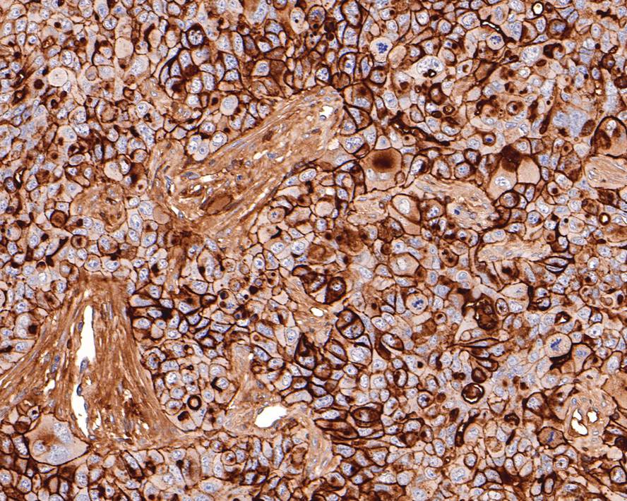
Immunohistochemical analysis of paraffin-embedded NCI-H441 xenograft tissue with Mouse anti-CD73 antibody (HA601011) at 1/600 dilution.
The section was pre-treated using heat mediated antigen retrieval with Tris-EDTA buffer (pH 9.0) for 20 minutes. The tissues were blocked in 1% BSA for 20 minutes at room temperature, washed with ddH2O and PBS, and then probed with the primary antibody (HA601011) at 1/600 dilution for 1 hour at room temperature. The detection was performed using an HRP conjugated compact polymer system. DAB was used as the chromogen. Tissues were counterstained with hematoxylin and mounted with DPX. -
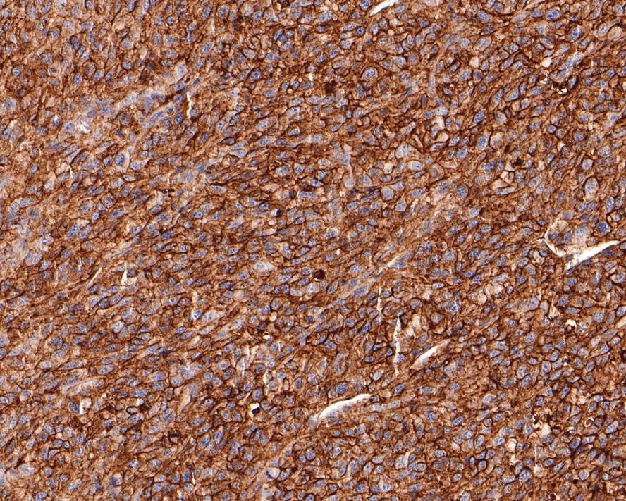
Immunohistochemical analysis of paraffin-embedded U87MG xenograft tissue with Mouse anti-CD73 antibody (HA601011) at 1/200 dilution.
The section was pre-treated using heat mediated antigen retrieval with Tris-EDTA buffer (pH 9.0) for 20 minutes. The tissues were blocked in 1% BSA for 20 minutes at room temperature, washed with ddH2O and PBS, and then probed with the primary antibody (HA601011) at 1/200 dilution for 1 hour at room temperature. The detection was performed using an HRP conjugated compact polymer system. DAB was used as the chromogen. Tissues were counterstained with hematoxylin and mounted with DPX. -
Immunocytochemistry analysis of U87-MG cells labeling CD73 with Mouse anti-CD73 antibody (HA601011) at 1/100 dilution.
Cells were fixed in 4% paraformaldehyde for 10 minutes at 37 ℃, blocked with 2% BSA for 30 minutes at room temperature. Cells were then incubated with Mouse anti-CD73 antibody (HA601011) at 1/100 dilution in 2% BSA overnight at 4 ℃. Goat Anti-Mouse IgG H&L (iFluor™ 488, HA1125) was used as the secondary antibody at 1/1,000 dilution. PBS instead of the primary antibody was used as the secondary antibody only control. Nuclear DNA was labelled in blue with DAPI. -
Flow cytometric analysis of U87-MG cells labeling CD73.
Cells were fixed and permeabilized.Then stained with the primary antibody (HA601011, 1ug/ml) (red) compared with Mouse IgG1 Isotype Control (green). After incubation of the primary antibody at +4℃ for an hour, the cells were stained with a iFluor™ 488 conjugate-Goat anti-Mouse IgG Secondary antibody (HA1125) at 1/1,000 dilution for 30 minutes at +4℃. Unlabelled sample was used as a control (cells without incubation with primary antibody; black).
Please note: All products are "FOR RESEARCH USE ONLY AND ARE NOT INTENDED FOR DIAGNOSTIC OR THERAPEUTIC USE"
同靶点&同通路的产品
CD73 Mouse Monoclonal Antibody [A6G1]
Application: WB,IHC-P,IF-Tissue,FC
Reactivity: Human
Conjugate: unconjugated
CD73 Mouse Monoclonal Antibody [6E-G3]
Application: WB,IHC-P,FC
Reactivity: Human
Conjugate: unconjugated
CD73 Recombinant Rabbit Monoclonal Antibody [JM11-40]
Application: WB,IHC-P,IF-Tissue
Reactivity: Human
Conjugate: unconjugated
CD73 Rabbit Polyclonal Antibody
Application: WB,IF-Cell,FC
Reactivity: Human,Mouse
Conjugate: unconjugated
CD73 Mouse Monoclonal Antibody [A6G2]
Application: WB,IHC-P,IF-Cell,FC
Reactivity: Human,Mouse,Rat
Conjugate: unconjugated
CD73 Recombinant Mouse Monoclonal Antibody [A6G2-R]
Application: WB,IHC-P,IF-Cell
Reactivity: Human,Mouse,Rat
Conjugate: unconjugated
